 |
| |
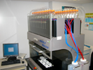
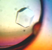
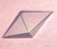
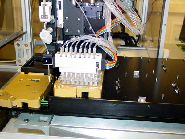
 With thousands of proteins and hundreds of different crystallization conditions each, it is essential to use automated crystallization and crystal visualization approaches. In the initial crystallization screen, we explore conditions that are incorporated into commercially available formulations and crystallization screens (Hampton Research, DeCode Genetics, Molecular Dimensions, NEXTAL, Jena Bioscience). With thousands of proteins and hundreds of different crystallization conditions each, it is essential to use automated crystallization and crystal visualization approaches. In the initial crystallization screen, we explore conditions that are incorporated into commercially available formulations and crystallization screens (Hampton Research, DeCode Genetics, Molecular Dimensions, NEXTAL, Jena Bioscience). |
|
| |
 | Strategy for protein crystallization | | Strategy for protein crystallization |
In an effort to reduce the number of crystallization conditions and maximize their effectiveness, we are collecting information about crystallization conditions, crystal morphology, space group, sequences and pI of protein, tags, and pH of crystallization. Similarly to other successful pilot projects our data are being analyzed for correlation with crystallization and structure determination success. Our initial results showed that the crystallization screens are redundant, and pointed to several less populated regions of crystallization space. Our most recent analysis suggests that large proteins (>60 kDa) and proteins with high pI crystallize less readily under the conditions that we use.
 Our analysis performed in 2007 of over 580 crystallization conditions showed that only about 300 conditions produce crystals. Based on this analysis, we have selected 288 out of 580 conditions for initial crystallization screens divided into three sets: CSH-96, CSM-96, and CSL-96 for high, medium, and low probability of obtaining crystals. Proteins that crystallize readily are revealed by the CSH-96 and CSM-96 (a total of 192 conditions), and only proteins that do not form macroscopic crystals are tested with an additional set of 192 conditions (CSL-96 and new development screen). These data are used similarly to approaches reported earlier to create more efficient, “tailor-made” crystallization screening kits.
New screens were designed (ANL-1 and ANL-2) that are now available via Qiagen.
We continue our effort to reduce the number of crystallization conditions and maximize their effectiveness.
Our analysis performed in 2007 of over 580 crystallization conditions showed that only about 300 conditions produce crystals. Based on this analysis, we have selected 288 out of 580 conditions for initial crystallization screens divided into three sets: CSH-96, CSM-96, and CSL-96 for high, medium, and low probability of obtaining crystals. Proteins that crystallize readily are revealed by the CSH-96 and CSM-96 (a total of 192 conditions), and only proteins that do not form macroscopic crystals are tested with an additional set of 192 conditions (CSL-96 and new development screen). These data are used similarly to approaches reported earlier to create more efficient, “tailor-made” crystallization screening kits.
New screens were designed (ANL-1 and ANL-2) that are now available via Qiagen.
We continue our effort to reduce the number of crystallization conditions and maximize their effectiveness.
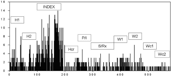
A recent survey of 2579 crystals (2009-10-01) showed that the crystallization screens are redundant, and pointed to several less populated regions of crystallization space (clearly seen in the PEGS-2 screen below).
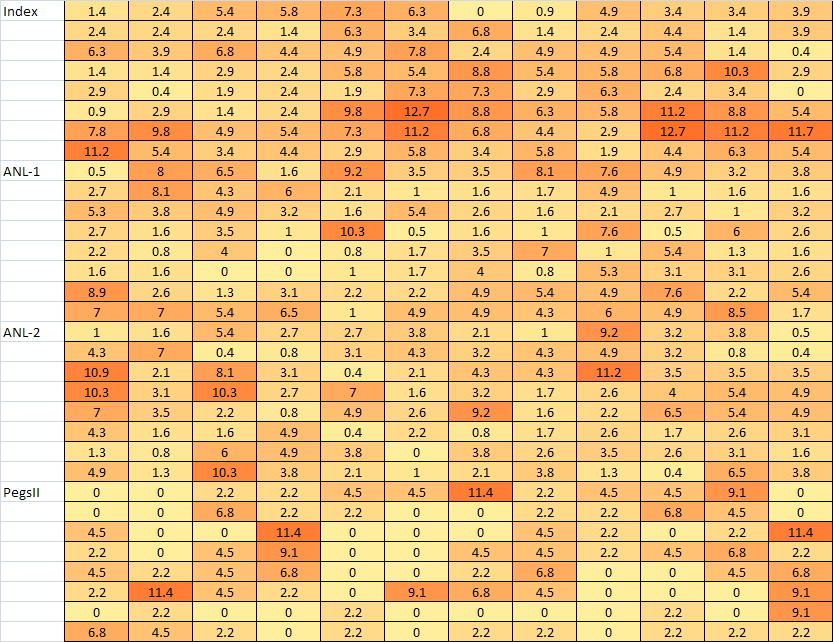
Although Index shows slightly higher success rate (0.47), ANL-1 and 2 screens (both show combined success rate 0.74) contain a more diverse set of conditions and they cover a larger crystallization space.
Based on the above analysis we have developed 4 new screens (MCSG1, MCSG2, MCSG3, and MCSG4) to include the most successful crystallization conditions observed previously (the Excel file with the conditions is available here: MCSG1-4 screen conditions).
Details: MCSG-I
MCSG-II
MCSG-III
MCSG-IV
The crystallization effort at the MCSG currently yields macroscopic crystals for 25% of purified proteins. Approximately one third of these crystals are already of data collection quality (are cryoprotected and have diffraction limits <3.2 Å, mosaicity <1°, suitable space group and unit cell, and no twinning). In all other cases (e.g., microcrystals, poor diffraction), we will perform optimization procedures to improve crystal quality. In optimization strategies, we vary the protein and precipitant concentration, the buffer concentration and pH, and the temperature and droplet size.
An important feature of our crystallization screening strategy is the inclusion of conditions that contain cryofreezing compounds. The addition of metals or other ligands is also known to affect protein crystallization. Moreover, these additives may remove heterogeneity associated with partial occupancy of ligands that may have been depleted during purification. Accordingly, we created two separate screens that include cofactors. Our metal screens contains FeCl2, MgCl2, MnCl2, CrCl3, CdCl2, ZnAcO2, CaCl2, CoCl2, CuCl2, WO4-2, PO4-2, and P2O7-3, and our organic cofactor screens include ATP, AMP, cAMP, GDP, NAD, NADP, Acetyl-CoA, and pyridoxal phosphate for ligand-directed crystallization. |
|
| |
| Production of x-ray-quality crystals and crystal optimization |
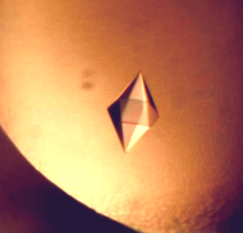
The overall goal of crystallization effort is to obtain x-ray-quality crystals for as many different proteins as possible. The crystallization effort at MCSG currently yields macroscopic crystals for 37.7% of purified proteins. In about half of the cases, crystals are large enough (at least 40-50 μm3) to be screened for diffraction properties at the synchrotron beamline. Approximately half of these crystals are already of data collection quality (are cryoprotected and have diffraction limits <3.2 Å, mosaicity <1°, suitable space group and unit cell, and no twinning). In all other cases (e.g., microcrystals, poor diffraction), we optimization procedures are performed to improve crystals quality. When the protein exhibits very strong crystallization polymorphism, up to five top conditions are optimized on the basis of crystal morphology and crystallization condition analyses.
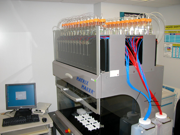
Optimization of crystallization conditions is one of the major bottlenecks of the structure determination pipeline and is one of the most challenging tasks. In optimization strategies, we vary the protein and precipitant concentration, the buffer concentration and pH, and the temperature and droplet size. We also experiment with additives (metal cofactor or/and organic cofactor screens) and different seeding methods. Crystal optimization is aided with a robotic solution maker and crystallization stations. We are using the 60-channel Matrix Maker (deCODE) to generate optimization and custom screens. The composition of solutions available on the Matrix Maker is set to cover about 90% of all successful crystallization conditions. Solutions not covered are prepared manually. For robot-assisted crystallizations, a 96-well format will used and for manual setups, a 24-well format will be used. Optimization screens are based on preliminary results and are developed with Crystal Monitor (deCODE) using three strategies:
- Component variation, a traditional grid optimization screen based on a single set of parent conditions.
- PBS cross (precipitant/buffer/salt), a combinatorial optimization screen based on two or more parent conditions. Precipitant, buffer, and salt from each of the selected conditions are recombined to produce all possible combinations of precipitant, buffer, and salt from the parent conditions.
- Cryo-additives, introduction of cryo-additives to crystallization conditions.
- Crystal optimization usually can be accomplished with fewer than 200 trials but may take 2-8 weeks. The total amount of protein required for optimization is on average 5-10 mg. A project may be abandoned when crystals do not appear to improve after a predefined set of conditions have been tried. When SeMet derivative of the protein demonstrates different crystallization properties, we may attempt microseeding with native crystals to produce SeMet-labeled protein crystals.
|
|
| |
| Cryofreezing crystals |
All x-ray diffraction data collection experiments are carried out at cryotemperatures, which requires screening for cryoconditions that do not affect x-ray diffraction properties. The optimal concentration of cryoprotectant is determined empirically and is monitored using x-ray diffraction. Several different approaches are being used to obtain conditions suitable for cryoprotection, such as growing crystals in the presence of cryoprotectant (glycerol, glycol, MPD, low-molecular-weight PEG, glucose, fructose, galactose, triose, isopropanol, and others), flash-washing crystals with cryoprotectant, or stabilizing crystals in gradually increasing concentrations of cryoprotectant. The simplest approach to obtaining crystals in a cryoprotectant is to grow the crystals in the cryoprotectant. With many examples in hand, we are now assembling a database of cryoprotectant concentrations for each commercial formulation.
Cryoprotected crystals are flash frozen in liquid nitrogen, gaseous nitrogen, or liquid propane. In our experience, the vast majority of protein crystals can be cryoprotected using some combination of these conditions. All data are collected at liquid nitrogen temperatures. For frozen protein crystals that do not show adequate diffraction properties, we either optimize the cryoconditions further, find a new crystal form, dehydrate the crystal or anneal the crystal.
|
|
| |
| Automated crystallization monitoring and documentation |
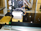
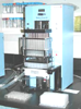
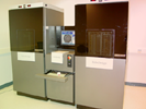
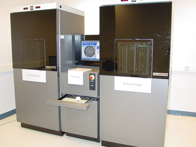
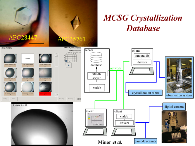
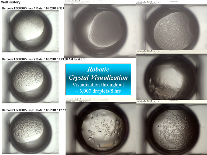
After careful analysis of the various systems on the market, we have elected to purchase a crystal storage and visualization system from Robodesign/Regaku. In this system, crystallization boxes are stored in two incubators that have a combined capacity to hold up to 800 bar coded SBS plates and 140 Linbro plates. Crystallization trials are imaged according to a preset schedule with a Minstral III microscope. The incubators and microscope are integrated and controlled by a single software package. All boxes are bar coded and data are maintained in the crystallization database using a terabyte server. Up to 16 users can access the data simultaneously. Currently, the crystallization robotic systems at the MCSG production sites can support up to 3,200 96-well plates and 560 24-well plates per year.
All crystallization images are recorded, stored, and analyzed. Each high-resolution image is approximately 4 Mbytes, In order to optimize storage capacity, an intelligent data management system is incorporated. The crystallization data from Robohotels are stored and managed by an Oracle database.
The manual crystallization trials are scored and archived in the crystallization database program developed at the MCSG’s University of Virginia facility. The system performs and records statistical analysis of crystallization trials, stores parameters of the crystallization experiment, images and annotates observations, and links them with a history of specific protein samples in a relational database. A bar code scanner is used to authenticate and identify users and quickly access previously labeled plates. A touch-screen monitor, located close to the stereo microscope, facilitates quick annotation of the observations. A digital camera mounted on the microscope records images, and the current drop image on the monitor may be compared directly with previous images.
Records from all crystallization stations are integrated with the central MCSG database (SGPDB). Both successes and failures will be documented, because both are important for a comprehensive analysis of the project.
|
|
| |
 Selected related publications: Selected related publications:
Kim Y, Dementieva I, Zhou M, Wu R, Lezondra L, Quartey P, Joachimiak G, Korolev O, Li H, Joachimiak A (2004) Automation of protein purification for structural genomics. J Struct Funct Genomics, 5, 111-8 Times cited: no data. [PubMed]
...
For a more exhaustive list of publications see the MCSG publications website. |
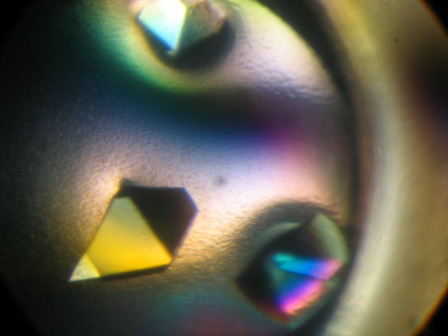 With thousands of proteins and hundreds of different crystallization conditions each, it is essential to use automated crystallization and crystal visualization approaches. In the initial crystallization screen, we explore conditions that are incorporated into commercially available formulations and crystallization screens (Hampton Research, DeCode Genetics, Molecular Dimensions, NEXTAL, Jena Bioscience).
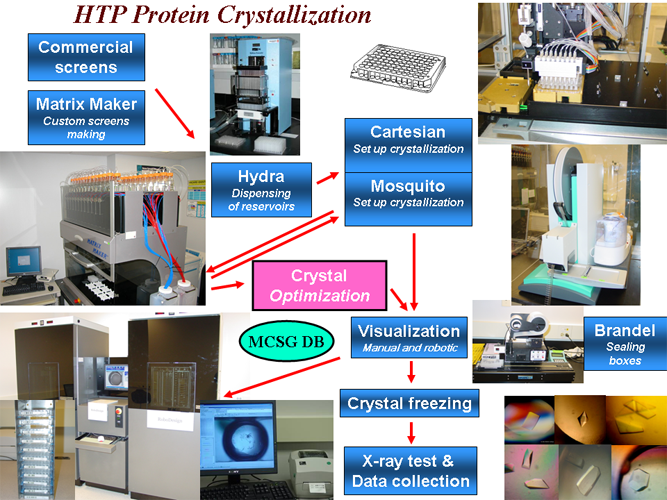


